Image analysis in inflammatory diseases

Psoriatic arthritis

Image data that can be used in the diagnosis of psoriatic arthritis: T1-weighted MRI (top left), fat-saturated MRI (top right), and fluorescence optical image series (bottom; left: early stage during the influx of the fluorescence agent, right: later stage during the influx of the fluorescence agent)
Exemplary visualization of the area scores resulting from the evaluation of fluorescence optical imaging in combination with the volume-registered fluorescence optical image series and the corresponding MRI of the hand. The left image is in the early stage of fluorescence agent influx, the right image was taken at a later stage.

Psoriatic arthritis (PsA) is a chronic inflammatory disease of the musculoskeletal system. Psoriatic arthritis is not curable. Currently, the progression of the disease can only be slowed down through therapy. Early diagnosis is therefore very important to minimize the symptoms for the patients. In addition, other arthritic diseases have similar symptoms but differ in treatment.

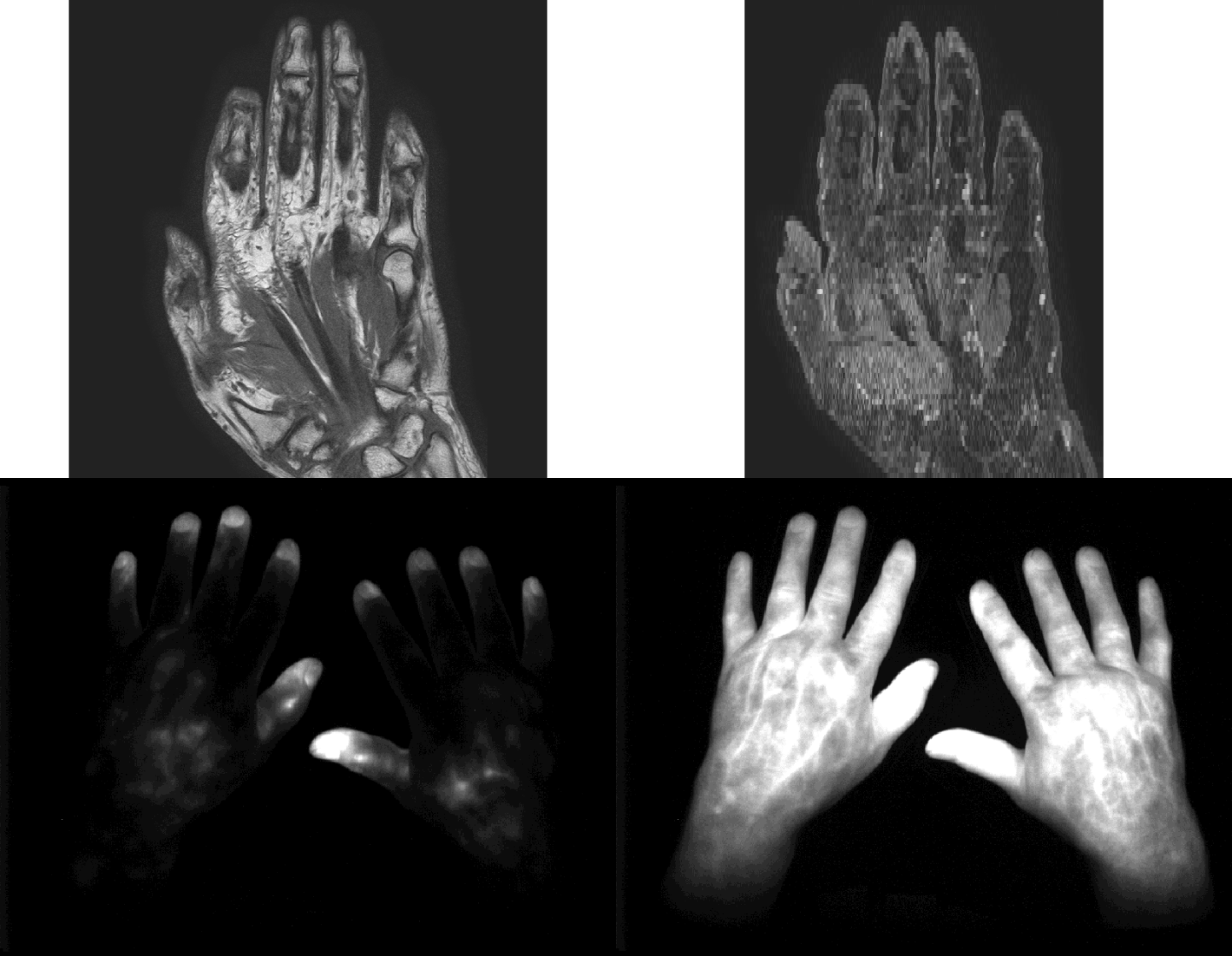
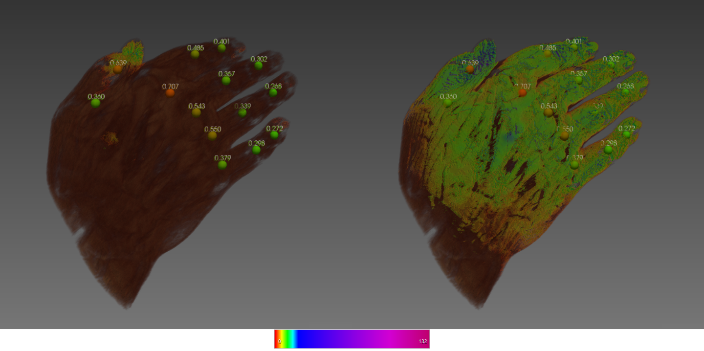
To support doctors in early detection, we are conducting research as part of the Fraunhofer "Cluster of Excellence Immune Mediated Diseases" to develop diagnostic solutions based on common imaging techniques. This includes the automated evaluation of X-ray images, ultrasound data, MRI, and fluorescence optical imaging. In the latter imaging technique, patients are injected with a so-called ICG dye, the distribution of which is then recorded in an image series. The distribution of the ICG is correlated with the activity of the metabolism in different areas. This allows conclusions to be drawn about potentially inflamed areas. These automated evaluations include, among other things, segmentations of individual bones in diagnosis-relevant regions, scoring of these regions, and diagnosis of existing symptoms. Since these evaluations are performed on different image modalities, we also deal with the non-rigid registration of the modalities. This enables a combined visualization of the results.

In addition, within the framework of the EU Innovative Medicines Initiative-funded project HIPPOCRATES, we collaborate with leading experts in Europe to improve the diagnosis, therapy of PsA, and the differentiation of other inflammatory diseases of the musculoskeletal tissue. This includes specialized imaging techniques such as High-Resolution Peripheral Quantitative Computed Tomography (HR-pQCT), in addition to the solutions mentioned above. Therapy successes should be made verifiable by recording and analyzing longitudinal data. In addition to the evaluation of image-based data, HIPPOCRATES also explores different cell parameters, so-called OMICS (proteomics, lipidomics, metabolomics, etc.), to generate an ideal rule set that correlates well with the progression of the disease.

Machine learning for image classification in histopathology

Automatically classified layers in the cross-sectional image of a 3D in vitro skin model

The Fraunhofer IGD has extensive capabilities in the analysis of histopathological image data. We rely on AI and deep learning methods to achieve precise and reliable results. We provide image analysis methods that enable automatic evaluation and detection of pathological changes in histopathological images.

As part of a project of the competence platform "New Drug Classes" of the Fraunhofer Cluster of Excellence for Immun-Mediated Diseases (CIMD), we have developed an AI-based automatic scoring method. This method automatically analyzes and evaluates images of H&E sections of 3D in vitro skin models. The Fraunhofer ISC in Würzburg develops these 3D in vitro skin models to test new drug classes. So far, a scoring method based on visual inspection of the skin section images by an expert has been used to evaluate the models (BSGC scoring).

Our developed method enables automated scoring of histopathological image data with high accuracy. This allows for the assessment of artificial skin models and stratification of fibrosis and other skin diseases in a short time without prior training of specialized personnel. In addition to direct classification, segmentation and classification of individual epidermis cell layers can be performed to obtain further information about the structure of the skin model. This makes it possible to identify interesting regions such as anomalies that may require further examination by experts. We use neural networks and rely on supervised learning to achieve high accuracy and robustness. The developed methods can also be applied to other skin diseases such as malignant melanoma or other use cases such as burn and wound models.

Further information

Healthcare Industry

We dedicate our services to companies in the industrial healthcare sector and medical care.